Home » Uncategories » Anatomy Muscles Pelvis / Exploring The Pelvis 3d Muscle Lab / To achieve both these tasks, the pelvic floor is composed of several overlapping sheets of muscles and connective tissues.
Anatomy Muscles Pelvis / Exploring The Pelvis 3d Muscle Lab / To achieve both these tasks, the pelvic floor is composed of several overlapping sheets of muscles and connective tissues.
Anatomy Muscles Pelvis / Exploring The Pelvis 3d Muscle Lab / To achieve both these tasks, the pelvic floor is composed of several overlapping sheets of muscles and connective tissues.. These muscles, including the gluteus maximus and the hamstrings, extend the thigh at the hip in support of the body's weight and propulsion. Ischiopubic ramus on either side. Muscles an important group of muscles in the pelvis is the pelvic floor. The muscles within the pelvis may be divided into two groups: These two muscles join each other and then attach to the lesser trochanter.
They have several functions, including helping to support the pelvic organs. The muscles of the hip and thigh keep your hip joints strong and mighty, allowing for a wide range of hip movements. Each hip bone consists of 3 parts. These two muscles join each other and then attach to the lesser trochanter. The bony pelvis comes together to provide support for the pelvic muscles and connective tissues, which, in turn, provide attachments and support for the pelvic organs.
The Pubococcygeal Muscle Pc Muscle And Attachments Yoganatomy from cdn-aolkg.nitrocdn.com The pelvic floor muscles include; The pelvic girdle (hip girdle) is formed by a single bone, the hip bone or coxal bone (coxal = hip), which serves as the attachment point for each lower limb. Now that you watched the video, you shou. Lower margin of pubic symphysis. Use the mouse scroll wheel to move the images up and down alternatively use the tiny arrows (>>) on both side of the image to move the images.>>) on both side of the image to move the images. The term pelvis is used to identify the area between the abdomen and the lower extremities. The pelvic floor has two inherently conflicting functions: Therefore, an appreciation of the female pelvic musculoskeletal anatomy is critical for understanding the pelvic support system.
The four groups are the anterior group, the posterior group, adductor group.
The function of the pelvic floor is to help assist with child birth, prevent incontinence and support organs within the pelvis. Arcus tendineus levator ani and the ischial spine Use the mouse scroll wheel to move the images up and down alternatively use the tiny arrows (>>) on both side of the image to move the images.>>) on both side of the image to move the images. These structures include the urinary system, genital organs, muscles, veins, nerves, arteries, and pelvic measurements. The other is to control the openings of the rectum and urogenital organs that pierce the pelvic floor and make it weaker. It is composed of three separate paired muscles; Together, these muscles straighten your knee, stabilize your knee joint, assist in flexing your hip (drawing your knee towards your chest), and help absorb force when you land after jumping or leaping. Attached to the pelvis are muscles of the buttocks, the lower back, and the thighs. The levator ani muscles are the largest group of muscles in the pelvis. They form a large sheet of skeletal muscle that is thicker in some areas than in others. Muscles that attach from the pelvis to the trunk and cross the lumbosacral joint muscles that attach from the pelvis to the thigh/leg and cross the hip joint pelvic floor muscles that are located wholly within the pelvis Large ligaments, tendons, and muscles around the hip joint hold the bones (ball and socket) in place and keep it from dislocating. The quad muscles— which form the meaty mass on the front of your thighs — are among your strongest muscle groups, and play a critical role in athletic activities.
These muscles origin in continuity from the body of the pubis, along a tendinous arch over the obturator internus fascia, and the ischial spine. Use the mouse scroll wheel to move the images up and down alternatively use the tiny arrows (>>) on both side of the image to move the images.>>) on both side of the image to move the images. To support the abdominal and pelvic viscera It is composed of three separate paired muscles; The floor of the pelvis is formed by the two muscles named levator ani and coccygeus.
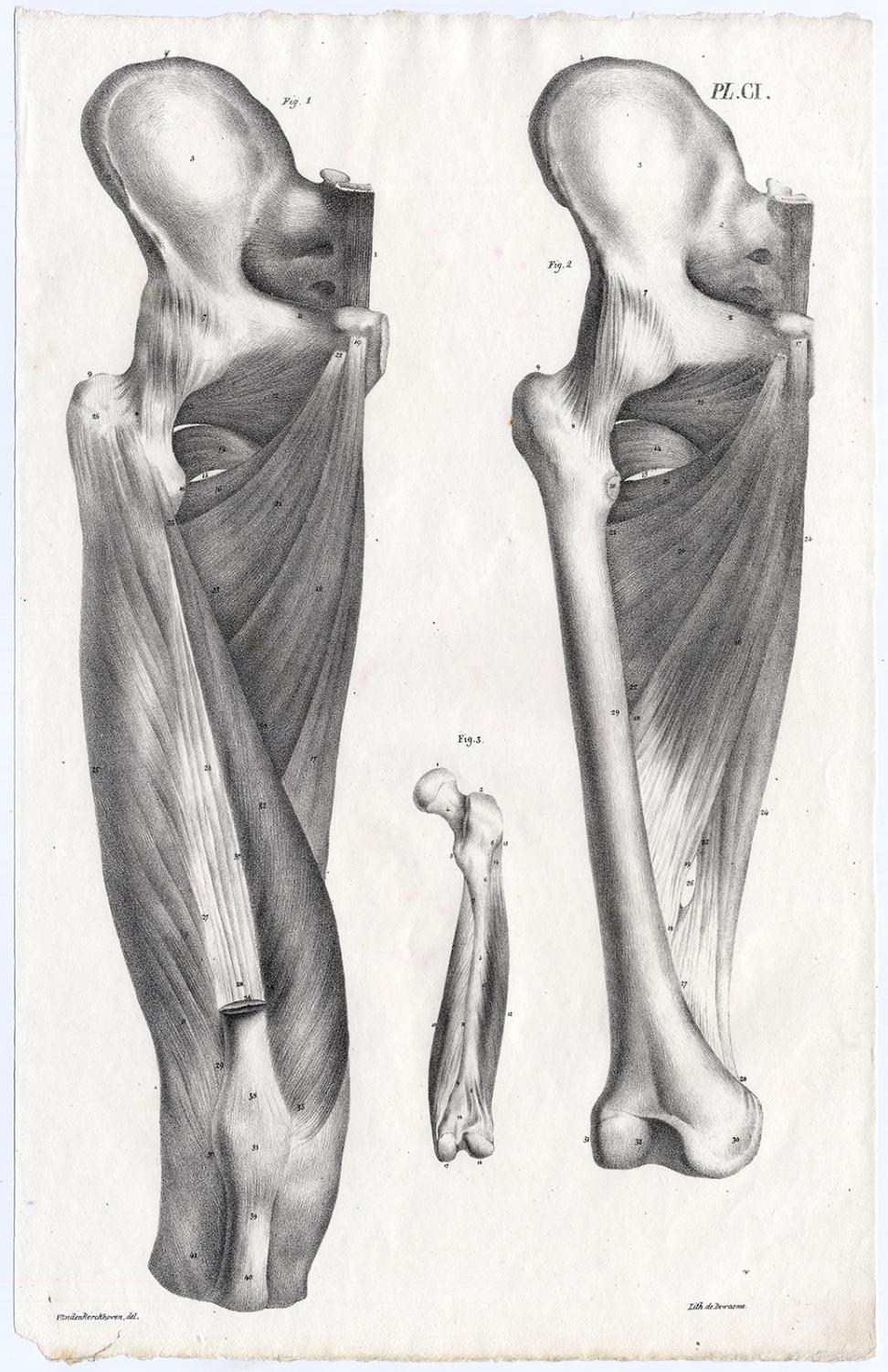
Antique Print Human Anatomy Muscles Upper Leg Femur Bony Pelvis Ci Cloquet 1821 Kunst Nbsp Nbsp Grafik Nbsp Nbsp Poster Theprintscollector from pictures.abebooks.com The muscles within the pelvis may be divided into two groups: The iliopsoas muscle consists of the iliac muscle, which comes from the inner surface of the ilium in the pelvis, and the psoas muscle, which originates from the vertebral column. (1) the obturator internus and the piriformis, which are muscles of the lower extremity, and will be described with these (pages 476 and 477); Muscles an important group of muscles in the pelvis is the pelvic floor. Together, these muscles straighten your knee, stabilize your knee joint, assist in flexing your hip (drawing your knee towards your chest), and help absorb force when you land after jumping or leaping. It can be described as one of the bodies diaphragms. Use the mouse scroll wheel to move the images up and down alternatively use the tiny arrows (>>) on both side of the image to move the images.>>) on both side of the image to move the images. The many muscles of the hip provide movement, strength, and stability to the hip joint and the bones of the hip and thigh.
Attached to the pelvis are muscles of the buttocks, the lower back, and the thighs.
They also help the anus function. The pelvic girdle (hip girdle) is formed by a single bone, the hip bone or coxal bone (coxal = hip), which serves as the attachment point for each lower limb. They form a large sheet of skeletal muscle that is thicker in some areas than in others. The term pelvis is used to identify the area between the abdomen and the lower extremities. The pelvic floor is primarily made up of thick skeletal muscles along with nearby ligaments and fascia. The floor of the pelvis is formed by the two muscles named levator ani and coccygeus. Pelvic outlet/ inferior aperture of pelvis separates pelvic cavity from perineum.its boundaries are as follows:. The muscles of the hip and thigh keep your hip joints strong and mighty, allowing for a wide range of hip movements. It is composed of three separate paired muscles; These two muscles join each other and then attach to the lesser trochanter. Large ligaments, tendons, and muscles around the hip joint hold the bones (ball and socket) in place and keep it from dislocating. The muscles within the pelvis may be divided into two groups: The 2 hip bones, along with the sacrum and coccyx of the spine form what's known as the pelvis.
Ischiopubic ramus on either side. The muscles within the pelvis may be divided into two groups: It is composed of three separate paired muscles; These two muscles join each other and then attach to the lesser trochanter. The pelvic floor has two inherently conflicting functions:
Medical Science Female Pelvis Model With Pelvic Floor Muscle Anatomy Model Buy Female Pelvis Model Pelvic Anatomy Model Medical Science Female Pelvis Model With Pelvic Floor Muscle Anatomy Model Product On Alibaba Com from sc04.alicdn.com The medial surface provides attachment for both transverse perinei, obturator internus and externus, piriformis, coccygeus and levator ani muscles. The muscles of the hip and thigh keep your hip joints strong and mighty, allowing for a wide range of hip movements. The bony pelvis comes together to provide support for the pelvic muscles and connective tissues, which, in turn, provide attachments and support for the pelvic organs. Enumerate the muscles of true pelvis. The other is to control the openings of the rectum and urogenital organs that pierce the pelvic floor and make it weaker. The pelvic floor muscles include; The ischium provides numerous points of attachment for pelvic and lower limb muscles. It's supplied by ventral rami of first and 2nd sacral nerves (s1, s2).
It's supplied by ventral rami of first and 2nd sacral nerves (s1, s2).
It is composed of three separate paired muscles; The pelvis's frame is made up of the bones of the pelvis, which connect the axial skeleton to the femurs, and therefore acts in weight bearing of the upper body. The levator ani is a broad sheet of muscle. It can be divided into the greater pelvis and the lesser pelvis. These two muscles join each other and then attach to the lesser trochanter. These muscles, including the gluteus maximus and the hamstrings, extend the thigh at the hip in support of the body's weight and propulsion. This mri male pelvis axial cross sectional anatomy tool is absolutely free to use. Piriformis the piriformis is a triangular muscle 1 on either side on the very front of the posterior wall of true pelvis. Arcus tendineus levator ani and the ischial spine These muscles origin in continuity from the body of the pubis, along a tendinous arch over the obturator internus fascia, and the ischial spine. These 3 parts actually start out as 3 separate bones at birth and fuse together throughout puberty. The pelvic floor has two inherently conflicting functions: (2) the levator ani and the coccygeus, which together form the pelvic diaphragm and are associated with the pelvic viscera.
0 Response to "Anatomy Muscles Pelvis / Exploring The Pelvis 3d Muscle Lab / To achieve both these tasks, the pelvic floor is composed of several overlapping sheets of muscles and connective tissues."


0 Response to "Anatomy Muscles Pelvis / Exploring The Pelvis 3d Muscle Lab / To achieve both these tasks, the pelvic floor is composed of several overlapping sheets of muscles and connective tissues."
Post a Comment